
It is a prevalent symptom impacting millions of people. Anything from the abdominal x-ray.

A Plain X Ray Of The Abdomen A Supine View B Erect View Showing A Download Scientific Diagram
Approach To The Abdominal X Ray Axr Undergraduate Diagnostic Imaging Fundamentals
Imaging Anatomy
After swallowing a barium solution X-ray films of the esophagus and stomach are taken.

Normal abdominal x ray. Here is his abdominal x-ray. Why is an MRI performed. Where the muscles and ligaments of the neck are forced to move outside their normal range.
The x-ray machine is positioned over the abdominal area. If you have mild uncomplicated antibiotic-associated diarrhea your bowel movements should gradually return to normal once your antibiotic treatment ends. The person will lie on a table or stand during the x ray.
A 33-year-old man presented repeatedly with severe abdominal pain and diarrhoea. An 82 year old man is admitted to the hospital for severe right flank pain. Inflammatory markers were mildly raised C reactive protein CRP 40 mg.
A neck X-ray also known as a. Abdominal MRI scans are used for. Because the alveoli are super-filled with air the patients lungs will typically appear darker than normal on chest X-ray because like we said air appears darker than tissue.
While clinical symptoms and confirmatory imaging Abdominal imaging with CT magnetic resonance transabdominal ultra-sonogram or endoscopic ultrasound can make a diagnosis of acute pancreatitis the most common presentation of AP is abdominal pain with elevated serum amylase and lipase usually three times the upper limit of normal. In this article we talk about the causes accompanying symptoms and. Abdominal Film X-Ray Medically reviewed by Stacy Sampson.
Sensations and intensity can vary and are subjective. A doctor can diagnose what is causing an abdominal mass with an MRI scan CT scan or X-ray. On chest X-ray this widened chest may be observed as well as flattening of the diaphragm due to the hyperinflated lungs pushing down on it.
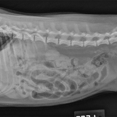
Tap onoff image to showhide findings. The psoas muscle edge is clearly defined on the left but not on the right. An unfortunate fact is that adhesions are unavoidable in surgery and the main treatment for adhesions is more surgery.
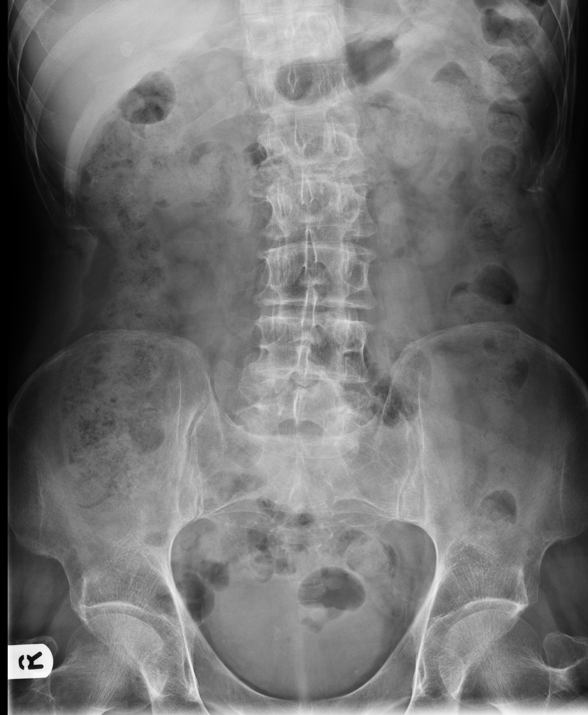
The small bowels mucosal folds are called valvulae conniventes and cross the full width of the bowel 5. Medical Dosimetry the official journal of the American Association of Medical Dosimetrists is the key source of information on new developments for the medical dosimetristPractical and comprehensive in coverage the journal features original contributions and review articles by medical dosimetrists oncologists physicists and radiation therapy technologists on clinical applications and. Normal - with faeces.
Abdominal pain is one of the most common symptoms seen by GPs. An x ray does not require anesthesia. Besides intestinal obstructions caused by adhesions that may be seen in an X-ray there are no diagnostic tests available to accurately diagnose an adhesion.
It may be the primary manifestation of respiratory cardiac neuromuscular psychogenic or systemic illnesses or a combination of these. A contrast medium usually a radiocontrast agent such as barium sulfate mixed with water is ingested or instilled into the gastrointestinal tract and X-rays are used to create radiographs of the regions of interest. Chest X-ray is also referred to as a chest radiograph chest roentgenogram or CXR.
Abdominal magnetic resonance imaging MRI is a noninvasive procedure that uses powerful magnets and radio waves to produce pictures of the inside of the. Hover onoff image to showhide findings. Chest-X-ray is a mainstay for follow-up in critically ill patients with covid-19 induced pneumonia.
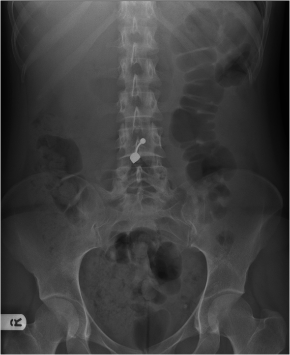
Renal colic was suspected and he was admitted for pain management. An x ray is performed at a hospital or an outpatient center by an x-ray technician and the images are interpreted by a radiologista doctor who specializes in medical imaging. There is no evidence of bowel obstruction or perforation.
Learn about abdomen x-ray abnormalities. A chest X-ray test is a very common non-invasive radiology test that produces an image of the chest and the internal organs. Dyspnea on exertion is a similar sensation.
Internal abdominal oblique Musculus obliquus internus abdominis Internal abdominal oblique is a broad thin muscular sheet found on the lateral side of the abdomenGoing from superficial to deep the external abdominal oblique internal abdominal oblique and transversus abdominis comprise the three distinct layers of the lateral abdominal wall. Irritable bowel syndrome IBS is a disorder in which the normal rhythmic movement of your gut bowel is disturbed - this can lead to abdominal pain bloating and excessive gas. Your doctor may order an abdominal MRI scan if you had abnormal results from an earlier test such as an X-ray CT scan or blood work.
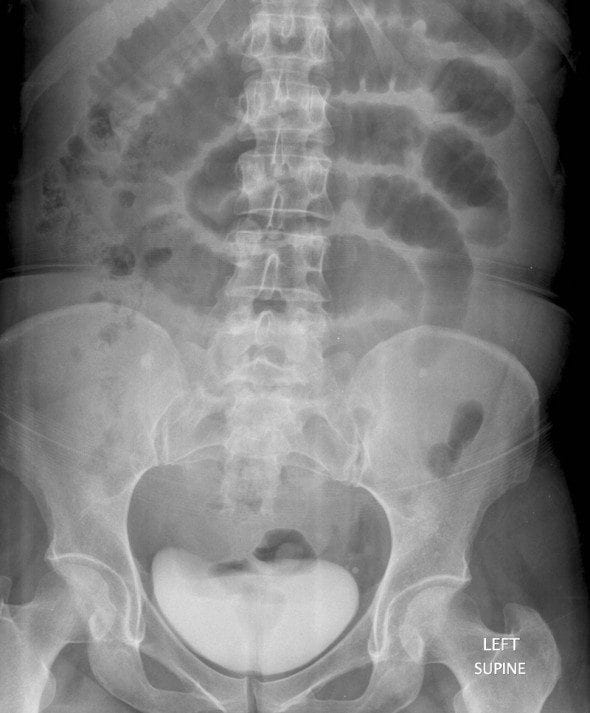
This is a normal abdominal X-ray with faecal material seen in the large bowel. De Barry et al. This testing usually involves checking one or more stool samples for the presence of a toxin made by C.
The X-ray is beamed to a special machine that converts it to a video and sends it to a TV-like monitor. Example of faeces and its typical mottled appearance 7. Questioning elicited an additional history of sore throat and mild dry cough.
This lets your radiologist follow the barium or iodine through your GI tract. Upper GI series with small bowel follow-through. Free gas under a diaphragm on an erect film indicates rupture of an abdominal hollow viscus such as the duodenum or small or large intestine.
Free gas - pneumoperitoneum - mimics of free intra-abdominal gas - pitfalls. Normal gas bubble on X-ray. A normal abdominal X-ray showing large bowel white arrow framing the small bowel black arrow 5.
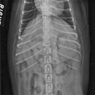
Yes it is the same patient 2. The chest radiograph also known as the chest x-ray or CXR is anecdotally thought to be the most frequently-performed radiological investigation globally although no published data is known to corroborate thisUK government statistical data from the NHS in England and Wales shows that the chest radiograph remains consistently the most frequently requested imaging test by GPs 2019 dataset 5. Is this lateral chest x-ray from the same patient as the last filmthe PA film.
Dyspnea also known as shortness of breath is a patients perceived difficulty to breathe. This can sometimes diagnose ulcers or other problems. Tutorial on abnormalities of the bowel gas pattern on abdominal X-ray.
To produce a chest X-ray test the chest is briefly exposed to radiation from an X-ray machine and an image is produced on a film or into a digital computer. Normal - with faeces. Normal Chest X-Ray and Approach to Chest X-Ray has been elaborately covered including the basics to the problem solving tools.
An upper gastrointestinal series also called a barium swallow or barium meal is a series of radiographs used to examine the gastrointestinal tract for abnormalities. Bone and soft tissue tumors of hip and pelvis.
Imaging Anatomy
Normal Abdomen X Ray Stock Image F003 3516 Science Photo Library

Axr Interpretation Litfl Ccc Investigations
Radiology Station Normal Abdomen X Ray With Anatomy Facebook


Abdomen Undergraduate Diagnostic Imaging Fundamentals
Normal Abdominal X Ray Radiology Case Radiopaedia Org
Abdominal X Ray Interpretation Axr Radiology Osce Geeky Medics

Abdominal X Ray Startradiology


